Tag Archives: cancer

Kidney cancer detected early with urine test

Nephrology_OncologyIf kidney cancer is diagnosed early – before it spreads – 80 percent of patients survive. However, finding it early has been among the disease’s greatest challenges.Now, researchers at Washington University School of Medicine in St. Louis have developed a noninvasive method to screen for kidney cancer that involves measuring the presence of proteins in the urine.The findings are reported March 19 in the journal JAMA Oncology.The researchers found that the protein biomarkers were more than 95 percent accurate in identifying early-stage kidney cancers. In addition, there were no false positives caused by non-cancerous kidney disease.

Read the rest of the article at http://www.medicalnewstoday.com/releases/291246.php.

Nanoparticles carry drugs selectively to lung tumor sites

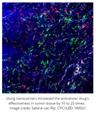
OncologyA new study published in the journal ACS Nano shows how nanoparticles might be used to fight lung cancer. The authors describe how they developed nanocarriers that can release chemotherapy drugs selectively at tumor sites without affecting tumor-free areas.The study is a project between scientists from two centers in Munich, Germany – the Helmholtz Zentrum München (HMGU) and the Ludwig-Maximilians-Universität (LMU) – and shows how the nanocarrier approach was able to deliver current cancermedicines for treating lung tumors more effectively than conventional methods.According to the World Health Organization, lung cancer is the leading cause of cancer death worldwide – accounting for 1.59 million deaths in 2012.Where cancer starts in the lung, it falls into one of two groups: small cell lung cancer or nonsmall cell lung cancer.Treatment of lung cancer depends on how big the cancer is and whether it has spread. Treatment options include surgery,chemotherapy, radiotherapy, or a combination of these.
Read the rest of the article at http://www.medicalnewstoday.com/articles/290462.php.

Sunlight continues to damage skin in the dark

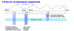
Dermatology_Radiology_OncologyMuch of the damage that ultraviolet radiation (UV) does to skin occurs hours after sun exposure, a team of Yale-led researchers concluded in a study that was published online by the journal Science.Exposure to UV light from the sun or from tanning beds can damage the DNA in melanocytes, the cells that make the melanin that gives skin its color. This damage is a major cause of skin cancer, the most common form of cancer in the United States. In the past, experts believed that melanin protected the skin by blocking harmful UV light. But there was also evidence from studies suggesting that melanin was associated with skin cell damage.In the current study, Douglas E. Brash, clinical professor of therapeutic radiology and dermatology at Yale School of Medical, and his co-authors first exposed mouse and human melanocyte cells to radiation from a UV lamp. The radiation caused a type of DNA damage known as a cyclobutane dimer (CPD), in which two DNA “letters” attach and bend the DNA, preventing the information it contains from being read correctly. To the researchers’ surprise, the melanocytes not only generated CPDs immediately but continued to do so hours after UV exposure ended. Cells without melanin generated CPDs only during the UV exposure.

Read the rest of the article at http://www.medicalnewstoday.com/releases/289728.php.

Early detection of heart attacks aided by gold nanoparticles

Cardiology_IM_FM_GP-Oncology_Dentistry_Hematology

NYU Polytechnic School of Engineering professors have been collaborating with researchers from Peking University on a new test strip that is demonstrating great potential for the early detection of certain heart attacks.

Kurt H. Becker, a professor in the Department of Applied Physics and the Department of Mechanical and Aerospace Engineering, and WeiDong Zhu, a research associate professor in the Department of Mechanical and Aerospace Engineering, are helping develop a new colloidal gold test strip for cardiac troponin I (cTn-I) detection. The new strip uses microplasma-generated gold nanoparticles (AuNPs) and shows much higher detection sensitivity than conventional test strips. The new cTn-I test is based on the specific immune-chemical reactions between antigen and antibody on immunochromatographic test strips using AuNPs.

Compared to AuNPs produced by traditional chemical methods, the surfaces of the gold nanoparticles generated by the microplasma-induced liquid chemical process attract more antibodies, which results in significantly higher detection sensitivity.

cTn-I is a specific marker for myocardial infarction. The cTn-I level in patients experiencing myocardial infarction is several thousand times higher than in healthy people. The early detection of cTn-I is therefore a key factor of heart attack diagnosis and therapy.

The use of microplasmas to generate AuNP is yet another application of the microplasma technology developed by Becker and Zhu. Microplasmas have been used successfully in dental applications (improved bonding, tooth whitening, root canal disinfection), biological decontamination (inactivation of microorganisms and biofilms), and disinfection and preservation of fresh fruits and vegetables.

The microplasma-assisted synthesis of AuNPs has great potential for other biomedical and therapeutic applications such as tumor detection, cancer imaging, drug delivery, and treatment of degenerative diseases such as Alzheimer’s.

The routine use of gold nanoparticles in therapy and disease detection in patients is still years away: longer for therapeutic applications and shorter for biosensors. The biggest hurdle to overcome is the fact that the synthesis of monodisperse, size-controlled gold nanoparticles, even using microplasmas, is still a costly, time-consuming, and labor-intensive process, which limits their use currently to small-scale clinical studies, Becker explained.

Adapted by MNT from original media release

http://www.medicalnewstoday.com/releases/288143.php


Animal-specific sugar may drive cancer risk in people who eat red meat

Oncology_Hematology-Oncology_Gastroenetrology

Reearchers at the University of California-San Diego School of Medicine report findings on the role a sugar specific to red meat may play in forming tumors in humans.


The researchers wanted to understand why people who eat a lot of red meat are at higher risk for certaincancers, while people who eat other types of meat are not.

The team first conducted a systematic survey of common foods and found that red meats – beef, pork and lamb – are rich in a sugar called Neu5Gc and provide the primary sources of this sugar in the human diet.

From previous studies, the researchers had found that Neu5Gc can be absorbed into human tissues.

From these findings, the team hypothesized that eating red meat could, therefore, promote potentially cancer-forming inflammation if the body is constantly generating antibodies against Neu5Gc, which is a foreign molecule.

In a mouse model engineered to have a deficiency of this sugar, the scientists found that feeding the mice Neu5Gc resulted in systemic inflammation, which was associated with a fivefold increase in spontaneous tumor formation.

Principal investigator Dr. Ajit Varki, distinguished professor of Medicine and Cellular and Molecular Medicine, and member of the UC-San Diego Moores Cancer Center, says:

“Until now, all of our evidence linking Neu5Gc to cancer was circumstantial or indirectly predicted from somewhat artificial experimental setups. This is the first time we have directly shown that mimicking the exact situation in humans – feeding non-human Neu5Gc and inducing anti-Neu5Gc antibodies – increases spontaneous cancers in mice.”

As the researchers did not expose the mice in the study to carcinogens or attempt to artificially induce cancers, they believe Neu5Gc is strongly associated with increased cancer risk.

Proving the association in humans will be ‘much harder’

“The final proof in humans will be much harder to come by,” Dr. Varki says. “But on a more general note, this work may also help explain potential connections of red meat consumption to other diseases exacerbated by chronic inflammation, such as atherosclerosis and type 2 diabetes.”

However, Dr. Varki also admits the team’s findings – which are published in the Proceedings of the National Academy of Sciences – represent a “catch-22,” as moderate amounts of red meat can be a source of good nutrition for young people. Dr. Varki hopes that the team’s future work will provide practical solutions to this.

In August, Medical News Today reported on a study suggesting that the increased risk of colorectal cancer associated with eating red meat may be reduced by consumption of resistant starch.

The researchers behind that study suggested, therefore, that by adding natural sources of resistant starch – bananas, beans, chickpeas, lentils, whole grains – to a diet high in red meat, the increased risk of colorectal cancer may be diminished.

Another study in 2014 also suggested that higher red meat intake during early adulthood was associated with a 22% increased risk for breast cancer.

That study found that replacing one portion of red meat a day with a portion of another high-protein food – for instance, legumes, poultry, nuts or fish – was associated with a 14% lower risk of breast cancer.

Written by David McNamee

http://www.medicalnewstoday.com/articles/287494.php


Ways to prevent and manage jaw bone disease highlighted 

Dentistry_Oncology

A review of more than a decade’s worth of research on osteonecrosis of the jaw–when the bone in the jaw is exposed and begins to starve from a lack of blood–points to an increased risk for patients taking certain drugs forosteoporosis, anticancer drugs or glucocorticoids, those undergoing dental surgery, and people with poor oral hygiene, chronic inflammation, diabetes, or ill-fitting dentures.

A number of prevention strategies may help protect at-risk individuals, and treatments that are available or under study include the use of antibiotics, surgery, teriparatide, bone marrow stem cell transplantation, and laser therapy.

“Patients need to maintain good oral hygiene especially if they are taking antiresorptive therapy in order to reduce their risk,” said Dr. Aliya Khan, lead author of the Journal of Bone and Mineral Research review.

http://www.medicalnewstoday.com/releases/287077.php


Thrombotic effects may delay immediate breast reconstruction after mastectomy

Oncology_Hematology-Oncology_Plastic Surgery

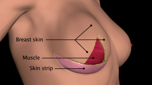
Immediate breast reconstruction following mastectomy is becoming more prevalent. However, in breast cancerpatients undergoing simultaneous chemotherapy, thrombotic complications can arise that can delay or significantly modify reconstructive plans. Outcomes of cases illustrating potential complications are published in the current issue of Annals of Medicine and Surgery.

Chemotherapy is increasingly used to treat larger operable or advanced breast cancer prior to surgery. Chemotherapy delivered via the placement of a central venous line that remains in place for the duration of treatment can result in pre-operative thromboembolic events, which can require the administration of anticoagulation agents. This in turn can complicate subsequent surgery and is particularly significant when complex reconstruction is anticipated immediately following the mastectomy.

“There is limited research on the impact of this complication on breast reconstruction pathways and guidance for optimal management of these patients,” notes Professor Charles M. Malata, FRCS (Plast), who is Consultant Plastic & Reconstructive Surgeon at the Cambridge Breast Unit (Cambridge University Hospitals NHS Foundation Trust) and Professor of Academic Plastic Surgery at the Postgraduate Medical Institute of Anglia Ruskin University. “My colleagues and I present our clinical experience over four years of patients with breast cancer who developed thrombotic complications of their neoadjuvant chemotherapy venous lines prior to mastectomy and immediate breast reconstruction.”

Investigators analyzed the pathways of seven breast cancer patients who had received primary chemotherapy during which they experienced pre-operative line-related thrombosis requiring anticoagulant therapy. Five of these patients were able to undergo surgeries as planned, however in two instances this was not the case.

Detailing the thromboembolic complications and the consequences for reconstruction, the investigators report that for one patient, surgery was delayed for three weeks while the thrombosis was managed, followed by two separate surgeries over a 12-month period to fully accomplish the reconstruction. In the second case, the reconstruction was achieved as planned, but only after a three-month delay and required anticoagulation therapy and pre-operative radiotherapy.

According to Professor Malata, “As these cases demonstrate, line-associated thrombosis will be increasingly encountered by surgeons as more patients receive neoadjuvant chemotherapy and are managed by indwelling venous access devices. With multidisciplinary cooperation among surgeons, oncologists, and hematologists, the surgical management plan for these patients can remain largely unaffected.” He further cautions, “Reconstructive surgeons should, however, be flexible enough to alter their surgical plan in patients who develop line-related thrombosis.”

http://www.medicalnewstoday.com/releases/286422.php


The secret life of anti-cancer drugs

Oncology

The public is bombarded with news of exciting developments in cancer research every day, with new anti-cancer drugs greeted with excitement.

But what happens to these drugs? When do they become accessible to cancer patients – if at all?

In a new review published in ecancermedicalscience, researchers trace the journey anti-cancer drugs take between discovery and clinical practice.

“Bringing a new medication to clinical practice is a lengthy process and involves many stakeholders with a very diverse background, ranging from researchers, regulatory agencies, payers, physicians and patients,” says Dr Felipe Ades of the Jules Bordet Institute, lead author of the review.

Prior to being incorporated into daily practice, drugs must obtain regulatory approval, succeed in changing the prescription habits of physicians, and ultimately gain the compliance of individual patients.

Each stage of development presents unique hurdles and challenges – for example, the average time for a drug to gain approval in the European Union is 418 days.

Once an anti-cancer drug is launched in the market, it takes around 3 years to have maximum impact on survival.

The complex life history of anti-cancer ends in the hands of the patient – who may not be inclined to take the drug at all.

“Aligning these different interests and expertise is a challenging task, but certainly a “win-win-win-win” situation,” Dr Ades says.

http://www.medicalnewstoday.com/releases/285564.php


Simple saliva test may reveal deadly diseases early enough to treat them

Dentistry_Oncology

UCLA research could lead to a simple saliva test capable of diagnosing – at an early stage – diabetes and cancer, and perhaps neurological disorders and autoimmune diseases.

The study, the most comprehensive analysis ever conducted of RNA molecules in human saliva, reveals that saliva contains many of the same disease-revealing molecules that are contained in blood. It was published online by the peer-reviewed journal Clinical Chemistry and will be published in the journal’s January 2015 special print issue, “Molecular Diagnostics: A Revolution in Progress.”

“If we can define the boundaries of molecular targets in saliva, then we can ask what the constituents in saliva are that can mark someone who has pre-diabetes or the early stages of oral cancer or pancreatic cancer – and we can utilize this knowledge for personalized medicine,” said Dr. David Wong, a senior author of the research and UCLA’s Felix and Mildred Yip Endowed Professor in Dentistry.

Wong said the test also holds promise for diagnosing Type 2 diabetes, gastric cancer and other diseases. “If you don’t look in saliva, you may miss important indicators of disease,” Wong said. “There seems to be treasure in saliva, which will surprise people.”

RNA, widely known as a cellular messenger that makes proteins and carries out DNA’s instructions to other parts of the cell, is now understood to perform sophisticated chemical reactions and is believed to perform an extraordinary number of other functions, at least some of which are unknown.

Wong’s research over the past decade has focused on identifying biomarkers in saliva. His laboratory discovered that some of the same RNA that is inside human cells are also present in saliva and can be used to detect diseases – a surprising finding, he said, because enzymes in saliva can degrade RNA, making the mouth “a hostile environment.”

The new research is a collaboration with Xinshu (Grace) Xiao, the paper’s other senior author and a UCLA associate professor of integrative biology and physiology. Using state-of-the-science genomics and bioinformatics, the researchers analyzed 165 million genetic sequences.

Among the many forms of RNA are some unusual ones that live in the mouth and in cells. For example, it wasn’t known until very recently that RNA comes in a circular form; the linear form has long been known. But the UCLA scientists identified more than 400 circular RNAs in human saliva – the first discovery of circular RNA in saliva or any body fluid – including 327 forms that were previously unknown.

Circular RNA’s function in saliva is not entirely understood, although it does serve as a sponge for tiny RNA molecules called microRNAs, which bind to it.

“Circular RNAs in saliva may be protecting other RNAs,” said Xiao, who also is a faculty member in the UCLA College and a member of UCLA’s Molecular Biology Institute. It’s likely, she said, that circular RNAs in saliva protect microRNAs from being degraded.

MicroRNAs, which once seemed to be little more than molecular noise, play important roles in many cell types, and have been implicated in cancers and other diseases, Xiao said. One microRNA can regulate hundreds of genes, she said.

The scientists compared microRNA levels in saliva to those in the blood and other body fluids, and found the levels of microRNA in blood and in saliva are very similar – indicating that a saliva sample would be a good measure of microRNAs in the body.

They also found that saliva contains another class of small RNAs, called piwi-interacting RNAs, or piRNAs, which are produced by stem cells, skin cells and germ cells. There are very few piRNAs in blood and most other body fluids, but Xiao’s analysis showed that piRNA are abundant in saliva. Although their function is not yet known, Xiao said they may protect the body from viral infection.

While most RNA molecules translate genetic code from DNA to make proteins, there is also a class called non-coding RNAs that does not.

“Saliva carries with it non-coding RNAs, microRNAs, piRNAs and circRNAs that are biomarkers for disease and health monitoring,” said Wong, who also is associate dean of research at the UCLA School of Dentistry. “Had we not done this collaboration, we would never know that non-coding RNAs, microRNAs, piRNAs and circRNAs exist in saliva.”

Their overriding conclusion is that saliva has tremendous medical and scientific value. In the not-too-distant future, dentists might be able to take saliva samples to analyze for a variety of diseases. And, Wong says, the research could lead to a new category of self-diagnostic devices. “This could indicate that wearable gear that informs you whether you have a disease – even before you have any symptoms – is almost here,” he said.

The scientists, both members of UCLA’s Jonsson Comprehensive Cancer Center, will continue their research, aiming to better understand the biology behind their findings, including the function of piRNAs.

“With a collaborator like Dr. Xiao, UCLA will continue to spearhead this science,” Wong said. “Now we have the capability and technology to reach deeper and study non-coding RNA, which has never been done.”

Adapted by MNT from original media release

http://www.medicalnewstoday.com/releases/284671.php

Picture courtesy to nutrihealthmagazine.com.


The quality of biopsy is directly linked to survival in patients with bladder cancer

Oncology_Urology

UCLA researchers have shown for the first time that the quality of diagnostic staging using biopsy in patients with bladder cancer is directly linked with survival, meaning those that don’t get optimal biopsies are more likely to die from their disease.

The two-year study found that about half of bladder cancer patients who were biopsied had insufficient material – meaning there was no bladder wall muscle retrieved – to accurately stage the cancer. Additionally, the UCLA research team found that a suboptimal biopsy and incorrect tumor staging was associated with a significant increase in deaths from bladder cancer, said study first author Dr. Karim Chamie, an assistant professor of urology and surgical director of the bladder cancer program at UCLA.

“These findings are very important because while patients know about the stage of their cancer, they rarely question the quality of the biopsy,” said Chamie, who also is a researcher at UCLA’s Jonsson Comprehensive Cancer Center. “We hope these findings will help empower patients to ask about the quality of their biopsy and, if it is suboptimal, then urge their doctors to repeat the biopsy prior to deciding on what type of treatment to prescribe.”

The findings were published in the early, online edition of the peer-reviewed journal Cancer. The study was conducted at UCLA and the Cancer Surveillance Program at USC.

Chamie said that many times, biopsies only take tissue from the inner lining of the bladder itself, ignoring the underlying muscle wall. If the cancer has started to invade, tissue from the muscle wall needs to be examined to determine the next course of treatment.

For the study, the UCLA team reviewed the medical records of every patient diagnosed with “non-invasive” – meaning the cancer had yet not spread into the muscle – bladder cancer during the years 2004 and 2005 across medical institutes in Los Angeles County. Biopsy and surgery reports were reviewed.

The team found that about half the time, the surgeon either did not do an adequate biopsy or the pathologist did not clearly state the extent of the cancer invasion. When patients had aggressive tumors and their surgeons and pathologists appropriately staged them, the chance of dying of bladder cancer at five years was 8 percent.

If the surgeon did an inadequate job staging them, but the pathologist alerted the physician of inadequate staging, the five-year mortality rate was 12 percent.

However, if the pathologist did not comment on the extent of the cancer invasion, then the five-year mortality was 19 percent.

“Appropriately staging patients with bladder cancer is a skill set that every urologist and pathologist should have in his/her armamentarium,” Chamie said. “We believe the next step is to change the staging system for bladder cancer to incorporate the quality of staging. Not all stage I cancers are alike. Some patients may have stage II cancer, but because the biopsy was insufficient, these patients were inaccurately staged and may be undertreated. I really do believe that one reason why we have yet to see significant improvement in bladder cancer survival over the last two decades may, in part, be attributed to inadequate staging.”

The study sample included 1,865 patients, 335 urologists and 278 pathologists from medical institutes across Los Angeles County. Muscle was reported as present in 972, or 52.1 percent, absent in 564, or 30.2 percent, and was not mentioned at all in 329, or 17.7 percent of initial pathology reports.

Barbara James, 77, of Three Rivers in Central California was diagnosed bladder cancer a year ago by an urologist in her area, who also performed a biopsy and later removed the cancer. However, the urologist waited four months before going back in to check to ensure all the cancer was removed, James said.

More cancer was found and the urologist wanted to go in and remove more.

“It made me really nervous. I felt like he didn’t know what he was doing,” she said. “I decided I needed to go somewhere else.”

A friend, a neurosurgeon at UCLA, referred James to Chamie, who performed a more thorough biopsy, which found that James had invasive bladder cancer that was missed in the first biopsy. As a result, her bladder had to be removed and a new bladder built for her using a portion of her intestine. She’s recovering and advises anyone with bladder cancer to go to a major medical center for biopsy and staging.

Every year, nearly 75,000 Americans are diagnosed with bladder cancer, the vast majority with non-invasive disease. More than 15,500 will die.

“The omission of muscle in the specimen or its mention in the pathology report in nearly half of all diagnostic resections is associated with increased mortality, particularly in patients with aggressive disease,” the study states. “Because urologists cannot reliably discern between aggressive and indolent disease, we recommend that patients with bladder cancer should undergo adequate muscle sampling at the time of biopsy.”

The study was funded by the National Cancer Institute, UCLA’s Jonsson Comprehensive Cancer Center, the Surveillance, Epidemiology, and End Results Program and the STOP Cancer Foundation.

http://www.medicalnewstoday.com/releases/284232.php